What is Hyperopia?

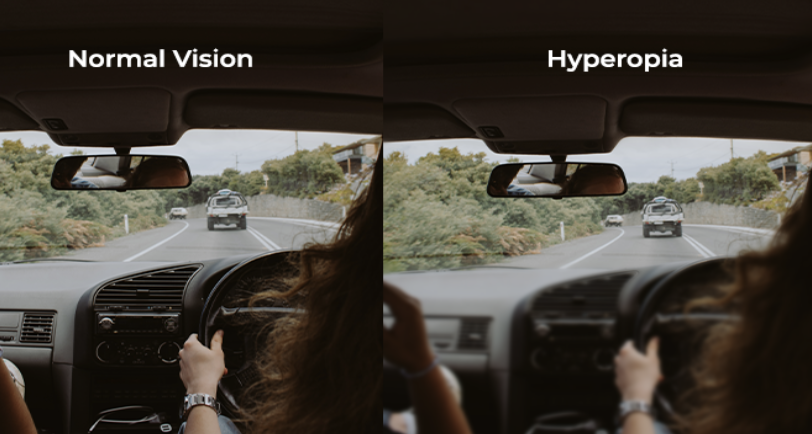
Hyperopia is also known as far-sightedness. It is a common type of refractive error that occurs when the light is focused behind the retina of the eye. Hyperopia affects near vision more, which means that distant objects can be seen more clearly than near objects.

There is a possibility that a combination of refractive errors can occur concurrently. For an example, hyperopia and astigmatism can occur in the same eye, or one eye can be short-sighted while the other is far-sighted. Hyperopia (rabun dekat in Malay) should not be confused with presbyopia (rabun tua in Malay) as they are not the same. Presbyopia is age-related and occurs when changes in the eye is less able to accommodate, and losing its ability to focus on close-up prints or objects. Hyperopia can occur at any age while presbyopia usually occurs over the age of 40.

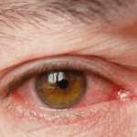
Symptoms of Hyperopia/Far-sightedness

  • Difficulty in seeing close-up objects
  • Eye strain or eye tiredness
  • Headaches, especially when doing close-up tasks
  • Blurred vision
  • Squinting
  • Fatigue

These symptoms tend to be more obvious after doing some near work such as reading, computer use, mobile phone activity and so on. The symptoms can vary, depending on the prescription. For individuals with mild farsightedness, the symptoms may not be so noticeable. Regular eye examinations are important to ensure that your vision is optimal. Individuals with moderate or severe hyperopia, may notice their vision is blurred for near and distant objects.

Causes of Hyperopia

Hyperopia occurs when images are focused behind the retina. From an anatomical standpoint, it is seen in eyes that are shorter. The cornea also plays a major role. If the cornea is too flat or less curved than normal, it might also cause images to fall behind the retina.

There is a genetic component involved with hyperopia, so most hyperopic individuals are born with a preponderance for it. Studies show the refractive errors are primarily influenced by multiple genes, so if a direct family member such as a parent or sibling is hyperopic, there is an increased likelihood of developing hyperopia. External factors such as lifestyle and diet may also influence changes.

Treatment

In young individuals, treatment may not be necessary because the natural lenses in the eyes are flexible enough to accommodate and compensate for mild to moderate degrees of hyperopia. The common management for hyperopic individuals is with glasses or contact lenses

  • Glasses -This is the most direct, simple and safe way to improve vision caused by hyperopia. There are different types of lenses such as those for single vision, bifocal lenses and progressive multifocal lenses.
  • Contact lenses. Soft clear lenses are worn on the eyes. They are available in a variety of materials and designs such as soft and Rigid Gas Permeable (RGP) lenses. They are available in monofocal or multifocal designs. There are contact lenses that correct hyperopia and astigmatism as well.

For individuals who prefer to be glasses-free and not rely on any glasses or contact lenses:

Orthokeratology lens (Ortho-K):

Ortho-K is the fitting of a customized rigid gas permeable (RGP) lens that is worn overnight. While the individual sleeps with the RGP lenses on, the lenses will gently reshape the front surface of the eye known as cornea. Clear vision can be achieved the following day upon removal of the lenses. Ortho-K can lenses only correct low degrees of hyperopia.

Refractive Lens Exchange (Cataract surgery):

This procedure involves removing the natural lens in the eye and implanting an artificial lens that corrects the hyperopia. This procedure usually applies to individuals who are above 40 years old, around the time when cataracts may start to develop and presbyopia sets in. Following a refractive lens exchange, individuals will not require cataract surgery in the future.

Laser Refractive Surgery

Laser surgery is done by reshaping the curvature of the cornea using a low energy laser machine. It is a good option for patients with hyperopia including PRK, TransPRK, and LASIK. TransPRK is a no touch ‘LASIK’ and bladeless treatment which is able to treat up to +6.00D of hyperopia.

Does poor diet affect your eyes?

Did you ever know what you consume or eat does not just affect your body shape, weight and health? In fact, an individual’s diet can also impact their eye health and cause some irreversible damages.

Does too much salt affect your eyes?

Salt is important and essential in almost every recipe to make it tasty, but did you know the high level of sodium can increase the risk of having high blood pressure. Salt will not only bloat our body and affect our body tissues, it will also lead to the restriction of blood flow to our eyes when the blood pressure is high.

Hypertension due to raised systemic blood pressure (BP) both diastolic and/or systolic is associated with stroke, cardiovascular, renal or ocular disease. Researchers have shown high blood pressure as a significant risk factor for glaucoma progression. Glaucoma is a common blinding disease which is due to damage to the optic nerve and raised intraocular pressure. Therefore, too much salt can increase overall blood pressure and lead to an increase of intraocular pressure.

Is it too sweet for the eyes?

Diabetes is a disease that occurs when the blood glucose levels are too high and people living with diabetes are at greater risk of developing eye problems. When the body does not produce enough insulin or does not produce any insulin, it will remain in the blood and not reach any cells causing an increase of glucose in the blood. Diabetes damages the blood vessels all over the body.

Diabetic retinopathy is the most common diabetic eye condition. It damages or changes your eyes by blocking the tiny blood vessels that go to the retina and lead to swelling or bleeding. In some serious conditions, the new blood vessels may grow on the surface of the retina. Diabetes retinopathy can appear in patients with either Type 1 or Type 2 and if the condition is left untreated, there is a chance to cause blindness.

The long term result of having diabetes can result in developing a cloudy substance in the lens which is known as cataract. Cataract develops for multiple reasons and most commonly is age-related whereas diabetes also increases the risk of getting cataract. The risk of developing cataract in diabetes patients is caused by the changes of tissues in the eyes that make up the eye’s natural lens.

In conclusion, poor diet control will be a risk for the health of your eyes. However, you can prevent and lower the risk of developing eye problems by just making a few changes to your diet. Having a healthy and well-balanced diet that includes lots of fruits, vegetables and lean proteins is important for most people to get the right nutrients for eye health. Those with vision problems and those with very restrictive diets should talk to an eye professional about the right foods to eat for their eye condition. An eye health professional can help you create a diet that suits you and reduce the chances of developing age-related eye diseases.

Bahaya Eyeliner dan Maskara Bagi Mata

Kelopak mata adalah lipatan kulit di atas dan di bawah mata. Pada kelopak mata terdapat bulu mata yang keduanya berfungsi melindungi mata dari cedera dan benda asing, cahaya berlebihan, serta membantu menyebarkan air mata yang diproduksi oleh kelenjar air mata.

Mengenakan maskara dan eyeliner adalah untuk menonjolkan keindahan mata Anda. Namun, mungkin tidak terlintas di pikiran Anda bahwa penggunaan riasan mata dapat menyebabkan timbulnya infeksi bakteri pada kelopak mata, yang kemudian dapat menyebabkan peradangan dan masalah penglihatan. Menurut FDA, peralatan rias aman dari kontaminasi saat Anda membelinya, tetapi setelah beberapa kali penggunaan, ada kemungkinan kuman di kulit Anda dapat menyebar dan berkembang biak pada peralatan rias tersebut. Oleh karena itu, Anda dianjurkan untuk mengganti alat rias Anda secara berkala dan ini sangat penting untuk mencegah terjadinya infeksi kuman.

Gejala Peradangan Mata

  • Mata merah dan berair
  • Mata terasa sakit dan bengkak
  • Sensitif terhadap cahaya
  • Blurred vision

Jenis-jenis Infeksi/Peradangan Mata

  • Peradangan mata luar – Bintitan adalah bisul kecil yang terjadi akibat infeksi bakteri pada akar bulu mata (folikel).
  • Peradangan mata luar – ‘Konjungtivitis’ adalah peradangan pada konjungtiva yang disebabkan oleh kuman seperti bakteri dan virus.
  • Peradangan kelopak mata – ‘Blepharitis’ adalah peradangan pada kelopak mata yang disebabkan oleh penyumbatan kelenjar minyak kecil di dekat pangkal bulu mata.
  • Peradangan di dalam mata – ‘Endophthalmitis’ adalah peradangan serius di bagian dalam mata yang disebabkan oleh kuman

Tidak dapat dipungkiri bahwa riasan dan wanita tidak dapat dipisahkan. Ini adalah rutinitas sehari-hari bagi sebagian besar wanita di seluruh dunia. Oleh karena itu, berikut adalah langkah sederhana dan penting bagi Anda untuk menghindari masalah mata yang tidak diinginkan.

Tips Menggunakan Riasan Mata dengan Aman

  1. Hindari menggunakan kosmetik yang sudah kedaluwarsa karena dapat meningkatkan risiko infeksi akibat terbentuknya jamur dan bakteri. Meskipun sebagian besar kosmetik mengandung bahan pengawet, bakteri tetap dapat terbentuk. Jika Anda tidak yakin tentang masa simpan suatu produk, Anda disarankan untuk membuangnya setelah 3 bulan penggunaan.
  2. Hapus riasan mata sebelum tidur. Hal ini penting untuk menghindari penumpukan riasan pada area kelopak mata yang dapat menyebabkan peradangan mata.
  3. Hindari pemakaian riasan pada tepian kelopak mata Anda. Tepi kelopak mata Anda (tepi kelopak mata, dekat tempat tumbuhnya bulu mata) mengandung kelenjar minyak (kelenjar meibom). Kelenjar ini menghasilkan komponen berminyak yang berfungsi menjaga kelembaban mata. Memakai riasan di bagian pinggir kelopak mata dapat menyebabkan kelenjar pada area tersebut tersumbat sehingga menyebabkan mata kering dan tidak nyaman.
  4. Hindari berbagi perlengkapan kosmetik. Berbagi peralatan rias berbahaya karena dapat meningkatkan risiko penyebaran infeksi yang dapat menyebabkan konjungtivitis.
  5. Bacalah label dan hindari bahan-bahan yang berbahaya. Sebelum membeli produk makeup, pastikan Anda membaca dan mengidentifikasi bahan-bahan yang digunakan. Ada beberapa bahan yang digunakan yang berbahaya bagi kulit dan dapat meningkatkan risiko kanker.

Kesimpulannya, menggunakan dan mempraktikkan teknik penghapus riasan yang salah dapat membahayakan mata Anda. Jika mata Anda mengalami kemerahan, nyeri, rasa tidak nyaman, atau penglihatan kabur, hentikan penggunaan riasan mata dan segera cari perawatan dari dokter mata.

Ketumbit: Perbedaan, Penyebab dan Pengobatan

Ketumbit adalah benjolan kecil pada kelopak mata. Ada dua jenis bintitan, yaitu bintitan (hordeolum eksternal) dan kalazion (hordeolum internal). Jadi, apa perbedaan antara keduanya? Bintitan adalah benjolan kecil akibat infeksi bakteri ‘staphylococcus aureus’ yang menyebabkan kelopak mata menjadi bengkak, merah, dan nyeri. Kalazion disebabkan oleh kelenjar bulu mata yang tersumbat dan menyebabkan pembengkakan, tetapi tidak menimbulkan rasa sakit. Namun, kalazion jarang menyebabkan pembengkakan pada seluruh kelopak mata.

Anda lebih berisiko terkena sariawan jika Anda:

  1. Sering mengucek mata tanpa membersihkan tangan terlebih dahulu
  2. Tidak membersihkan kelopak mata dan bulu mata, terutama setelah menggunakan kosmetik
  3. Blefaritis, sejenis peradangan di sekitar tepi kelopak mata
  4. Memiliki masalah kesehatan terutama penderita diabetes

Bagaimana cara menyembuhkan sembelit?

Biasanya, ketumbit akan hilang dengan sendiri tanpa memerlukan rawatan. Bisul juga dapat diobati di rumah dengan mengompres hangat bisul beberapa kali sehari. Jika tidak sembuh, antibiotik akan diberikan terlebih dahulu. Namun, pada kasus yang lebih parah, operasi kecil mungkin diperlukan untuk mengobati abses. Perlu diketahui pula, sembelit dapat terjadi berulang kali. Oleh karena itu, menjaga kebersihan area mata, seperti kelopak mata dan bulu mata, sangat penting untuk mencegah timbulnya bintitan.

Metode kompres hangat adalah:

  1. Menggunakan sapu tangan atau kain bersih
  2. Rendam dalam air semi-panas dan peras hingga tidak ada air yang menetes.
  3. Tempelkan pada kelopak mata dengan mata tertutup selama 10 menit.
  4. Ulangi 2-3 kali sehari sesuai anjuran dokter Anda.

Cara membersihkan bulu mata

  1. Campurkan air bersih dan beberapa tetes sampo bayi dalam wadah kecil.
  2. Celupkan kapas ke dalam campuran tersebut lalu gosok perlahan kelopak mata dengan kapas tersebut. Hindari kontak dengan kornea.
  3. Lakukan 2-3 kali sehari
  4. Jika kelopak mata Anda terlalu kotor dan bersisik, oleskan air hangat terlebih dahulu agar kulit yang bersisik menjadi lembut dan mudah dibersihkan.

Meskipun katarak tidak berbahaya bagi penglihatan, namun dapat menyebabkan ketidaknyamanan dan memengaruhi penampilan Anda, yang dapat mengganggu aktivitas sehari-hari. Oleh karena itu, apabila kondisi tidak kunjung membaik setelah beberapa hari atau seminggu, segera periksakan diri ke dokter untuk mendapatkan penanganan lebih lanjut.

Differences: Pinguecula vs Pterygium

Pterygium and pinguecula are conjunctival growths that form on the surface of the eye. Both are not dangerous and will not cause blindness. In the early stage, there is no effect on vision and the main concern is cosmetic appearance, So what’s the difference between them?

Pterygium is the fleshy triangular mass that extends to the cornea while pinguecula is the yellowish elevated bump or lump that does not grow across the cornea. Pterygium can be a lot more obvious as it grows onto the clear surface of the cornea. It is also known to change the shape of the cornea causing changes in vision which is an increase in astigmatism. Pterygium may grow over a period of months to years until it covers the cornea, only then it will interfere with vision. Pinguecula usually develops on the side of the eye and doesn’t typically grow to a bigger size. Pinguecula does not typically harm your vision.

What causes these growths?

Both are caused by extended exposure to ultraviolet (UV) light (sunlight), wind and dust. They occur in people who spend more time outdoors, especially in hot, dry and sunny climates. The risk is also increased by not wearing sunglasses or a hat. People whose jobs expose them to UV light such as fishermen, farmers and contractors are also prone to develop this eye condition. Sailors, surfers and skiers are also more prone to develop pterygium as they encounter high levels of reflected UV light.

What do you feel?

Most people don’t feel anything. However, some people will experience foreign body sensation, grittiness and irritation in their eyes due to interference with the tear film. These symptoms are most difficult for contact lens wearers as the contact lens may not fit very well on the eye due to the uneven eye surface. Pinguecula and pterygium may occasionally get inflamed, and this will cause more discomfort, redness and grittiness. Furthermore, if the pterygium extends to the cornea, the curvature of the cornea will change and will cause some visual disturbance such as glare and blurred vision.

How are pterygium and pinguecula treated?

Once either one has formed on the eye, they will not go away by itself. An Ophthalmologist will do a full eye examination and determine the appropriate management for the pterygium and pinguecula. Lubricant eye drops can be prescribed to reduce irritation and discomfort and provide relief. Pterygium can remain small or will grow until extending towards the pupil.

It is important to have regular eye exams to monitor the progression of the pterygium. If eye drops do not help relieve symptoms and when a pterygium is large enough to interfere with vision, a pterygium removal surgery may be necessary to help restore vision. Unlike pterygium, pinguecula will not grow across your cornea, therefore surgery is not usually necessary. However, in some cases pinguecula can develop into a pterygium and affect vision.

How to prevent it?

Polarized sunglasses or hats with wide brim when outdoors can help to prevent and slow the progression of these types of tissue growth. People that work under exposure to sunlight can use appropriate protective eye equipment in work environments. Try to avoid exposure to environmental irritants such as smoke, dust, wind, and chemical pollutants. Protect your eyes from dust by wearing spectacles or goggles. It is also recommended to use artificial tears if you are exposed to dry conditions.

Mata berkedip sebelah – Normal atau Tidak?

Pernahkah Anda mengalami mengedipkan mata sendiri? Berkedip berulang kali dan tak terkendali. Biasanya terjadi pada kelopak mata atas, tetapi beberapa orang juga mengalaminya pada kelopak mata bawah. Kedutan mata atau dalam bahasa ilmiahnya myokimia adalah kejang otot pada kelopak mata. Biasanya berlangsung beberapa detik dan akan hilang dengan sendirinya. Kondisi ini tidak menyakitkan atau berbahaya, namun menganggu dan menimbulkan rasa tidak nyaman bagi yang mengalaminya. Namun, masalah ini mungkin merupakan tanda awal penyakit kronis jika kejang kelopak mata melibatkan jangka waktu lama atau gerakan tidak terkendali pada bagian wajah lainnya.

Diantara penyebab mata berkedut (eye twitching)

  • Stres dan kelelahan mental
  • Asupan kafein/alkohol
  • Dry Eye
  • Melihat layar komputer/ponsel/buku terlalu lama

Terkadang kedipan mata yang terus-menerus bisa menjadi tanda adanya masalah yang lebih serius seperti:

  • Peradangan kelopak mata (Blepharitis)
  • Kejang otot mata (Blepharospasm)
  • Kejang otot wajah (Facial spasm)

Namun, beberapa individu mengalami efek samping akibat masalah ini. Diantaranya adalah:

  • Kelopak mata terkulai (Ptosis)
  • Penglihatan ganda (Diplopia)
  • Blurred vision
  • Mata berair berlebihan

Oleh karena itu, jika Anda mengalami efek samping seperti ini, segeralah mencari penanganan dari dokter spesialis mata.

Cara mengatasi kedutan mata (eye twitching)

Kedutan mata biasanya hilang dengan sendirinya tanpa pengobatan. Anda dapat mencegah hal ini terjadi lebih sering dengan mengatasi penyebab masalah ini seperti:

  1. Mendapatkan istirahat dan tidur yang cukup
  2. Kurangi asupan kafein/alkohol
  3. Minum banyak air, tingkatkan frekuensi berkedip dan gunakan tetes air mata buatan jika perlu untuk masalah mata kering.
  4. Batasi waktu yang dihabiskan menggunakan komputer, telepon, atau membaca.

Jika blefarospasme terjadi, perawatan suntikan toksin botulinum (botox) dapat digunakan untuk meredakan kejang. Sementara itu, untuk kasus blefarospasme yang parah, pembedahan akan dilakukan.

What is digital eye strain (DES)?

Digital eye strain also known as computer vision syndrome, is a condition where a person experiences a temporary eye discomfort after prolonged use of electronic devices such as computer, tablet, mobile devices. The level of discomfort increases with the amount of digital screen use. DES can occur differently in each individual based on their visual device lifestyle, but their symptoms are usually the same.

Symptoms of DES

The most common symptoms associated with digital eye strain are

Causes of DES

  1. Reduction of blinking when staring at electronic devices is one of the reasons people experience DES. A person normally will blink about 15 times per minute. However, the blinking rate can be less than half when a person continuously stares at the screen.
  2. Glare can occur on devices with glass screens which contribute to DES due to the reflection and brightness of the screen.
  3. Improper viewing distance by staring at the computer at a too close or too far distance for a long time can cause DES.
  4. Poor seating position while staring on screen at a different height level can lead to many problems such as headache and neck and shoulder pain. This is because the position of the eye viewing is not parallel to the screen.
  5. Uncorrected vision problems such as short-sightedness, astigmatism, presbyopia and underlying eye problems can easily cause a person to be more symptomatic towards DES.
  6. Contact lens wear can increase the chance of having dry eyes and irritation.

Treatment & Prevention of DES

  1. Taking a break frequently while looking at a screen for long hours is very important. By practicing the “20-20-20” rule, for every 20 minutes look away from the screen and look at an object 20 feet away for at least 20 seconds. This allows the eyes to rest and refocus themselves.
  2. Reduce glare by adjusting the brightness on the screen or dimming the light near the screen can help to reduce the DES. An additional matte screen filter on top of the screen can also help.
  3. Ergonomics are crucial in the working environment.
    a) Adjusting the seating position while looking at the screen for long hours is important to prevent neck strain and shoulder pain while working. The chairs must be comfortably padded and conform to the body and the chair height should be adjusted so the feet rest flat on the floor. Arms have to be adjusted to provide support while typing and wrists shouldn’t rest on the keyboard when typing.
    b) Location of the computer screen should be 15 to 20 degrees below eye level (about 4 or 5 inches) as measured from the center of the screen and 20 to 28 inches from the eyes. Most people find it more comfortable by viewing the computer with their eyes looking downward.
    c) The arrangement of reference materials has to be below the monitor and above the keyboard. Otherwise, a document holder can be placed beside the monitor. The reason for positioning the documents is to reduce head effort to reposition from the document to the screen.
  4. Correct refractive errors and underlying eye problem by visiting an optometrist or ophthalmologist for proper management and eye prescription in order to have a better vision performing daily tasks.
  5. Contact lenses wearers can consider having a pair of glasses while staring at the devices for long periods especially during working hours. Using artificial tears to lubricate the eyes with or without contact lenses can reduce symptoms.

Digital eye strain can be diagnosed through a comprehensive eye examination. During the exam, it is important to tell your Ophthalmologist how often you use a computer or digital device at home or work. Generally an eye strain can be treated with simple methods, however prolonged eye strain and discomfort may be a sign of something serious that should be treated by an Ophthalmologist. Those living in areas such as Shah Alam, Cheras, Puchong, Shah Alam, Petaling Jaya, and Kepong can come visit our ophthalmologists to have their eyes examined.

Darurat Mata Merah

Most red eyes are usually not sight threatening and can recover without leaving any permanent damage to the eye.

However, one of the red eyes that may require immediate attention is Acute angle closure Glaucoma. Acute angle closure Glaucoma is vision or sight threatening and needs urgent and timely treatment to prevent irreversible damage to the optic nerve. Optic nerve is the “cable” that connects the eye to the brain and once it is damaged it cannot regenerate.

Some symptoms of acute angle closure glaucoma besides a red eye are usually headaches (in very severe cases nausea and vomiting), pain in the eye associated with blurring of vision and sometimes seeing haloes (due to swelling of the cornea due to increased intraocular pressure).

Acute angle closure glaucoma, is a true ocular emergency and needs to be treated urgently to prevent irreversible damage to the optic nerve.

The first thing to do is to reduce the intraocular pressure (IOP) with topical/oral anti-glaucoma medications. In certain cases, patients may require intravenous (IV) medications too. The doctor will also treat the inflammation with steroid eye drops.

Laser peripheral iridotomy (LPI) needs to be done as soon as possible to the affected eye and also for the other eye to prevent a similar problem in the future. Iridotomy is a procedure where we create a hole in the iris (with laser). If it not possible, then it has to be created surgically, to relieve the pupillary block due to angle closure glaucoma.

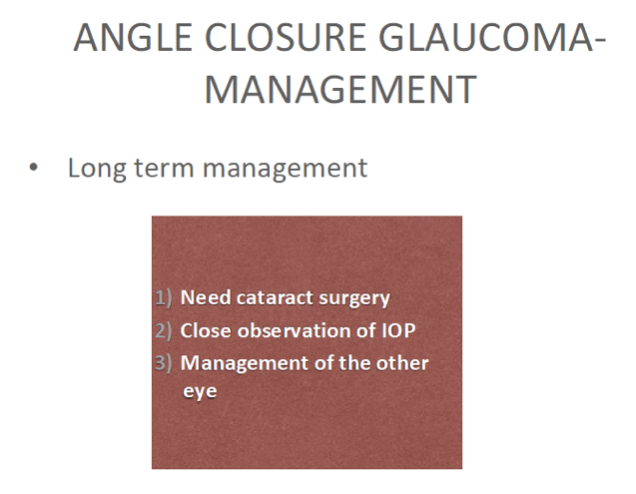
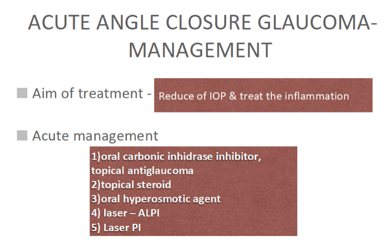
Once the IOP is controlled with laser treatment and anti-glaucoma medications and the affected eye has stabilized, there is a need to plan for cataract surgery in the future. All these are done to reduce and prevent irreversible damage to the optic nerve and thus maintain quality of life.

Watery Eyes

 Watery eyes or tearing is a condition when a person produces an excess of tears and there is an overflow of tears onto the face. Tears are produced from the tear gland known as lacrimal gland and they serve few important roles to the body. They help to wash away foreign particles and dust and also act as a component of the immune system that protects us from infection. Tears also help to moisture and lubricate our eyes. An overflow of tears or too much of it on the surface of the eye can make it difficult to see.

Tears drain from the eye through the tear ducts into the nose. A blocked tear duct or a narrow duct is the most common cause of watery eyes in adults. If the tear ducts are narrowed or blocked due to inflammation or infection, the tears will not be able to drain away and thus cause excessive tearing. Some other eye conditions can also cause watery eyes. Bumps on the eyelids like stye or chalazion which are caused by infection or inflammation along your eyelids can also prompt watery eyes. When eyelashes grow inwards toward your eyes, it rubs against the cornea and causes irritation. This condition known as ingrown eyelashes or trichiasis can cause watery eyes as well.

It is also common to temporarily produce excess tears when you are in a dry environment. When you stay long in a dry environment, it prompts your tear glands to produce and release more tears which is known as reflex tearing. Exposure to pollen, pet dander, mites and fumes can cause your eyes to turn red, itchy and watery which are known as allergies. Another cause of dryness is prolonged or over wear contact lenses which can increase tear evaporation. This will evoke your tear glands into overproduction of tears and result in watery eyes. Staring at your computer screen for a long time will also result in watery eyes. When focusing too much on a computer screen, it tends to reduce our blinking rate and lead to dryness. To avoid this, we can practice the 20-20-20 rule which means that for every 20 minutes, focus on an object 20 feet away for 20 seconds. This simple action can give your eye some rest.

Watery eye treatment depends on how severe the problem is and its cause. For blocked tear ducts, surgery can create a new channel from the tear sac to the inside of the nose which allows tears to bypass the blocked part of the duct. A visit to an Ophthalmologist can help in resolving a long-standing watery eye problem.

Dr Tai Lai Yong
Consultant Ophthalmologist
Oculoplastic, Orbital & Lacrimal Surgeon

Diabetes causes blindness

Diabetes is a metabolic disease that causes high blood sugar. The hormone insulin moves sugar from the blood into your cells to be stored or used for energy. Diabetes can happen when your body either doesn’t make enough insulin or can’t effectively use the insulin it makes. Untreated high blood sugar from diabetes can damage your eyes. People with diabetes can have an eye disease called diabetic retinopathy. This happens when high blood sugar levels damage blood vessels in the retina. These blood vessels will be blocked and leaky. Sometimes abnormal new blood vessels will grow on the retina. All of these changes can make your vision become worse.

Types of diabetic retinopathy

  1. Non-proliferative diabetic retinopathy is the early stage of the disease. The blood vessels in the retina are weakened and there may be fluid leaking into the retina which may lead to swelling of the retina.
  2. Proliferative diabetic retinopathy is the advanced stage of the disease. The blood vessels might rupture and the blood may leak into the vitreous cavity, this causes patients to have acute blurry vision.

Artificial intelligence (AI) fastest way in detecting diabetic retinopathy

Previously, diabetic retinopathy was analysed and graded manually, therefore it usually takes around an hour to identify the findings. Next, these patients diagnosed as diabetic retinopathy will go through a second round of grading to ensure that the diagnosis is accurate. Artificial intelligence(AI) programme is a new technology which can deliver diagnosis within just a couple of minutes and the accuracy for AI systems is about 90% because it eliminates human inconsistencies when analysing data.

How does artificial intelligence (AI) work?

It uses selena+ software which looks out for symptoms such as bleeding,swelling and microaneurysms, which would appear as tiny red lesions in retina photography. After uploading the retinal image, the software will compare it against the dataset it has been trained on, which comprises nearly 500,000 retinal images of patients with the diseases.

OasisEye Specialists is the first Eye Centre to initiate the use of Artificial Intelligence (AI) for diabetic eye screening in Malaysia. OasisEye is ahead in providing premier eye care to its patients and collecting data to further strengthen the current AI algorithms or develop new ones.

How to prevent blindness

Diabetic retinopathy is asymptomatic at an early stage. Therefore, patients with diabetic are advised to have an annual eye screening. Early detection and treatment can prevent vision loss.