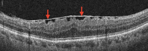
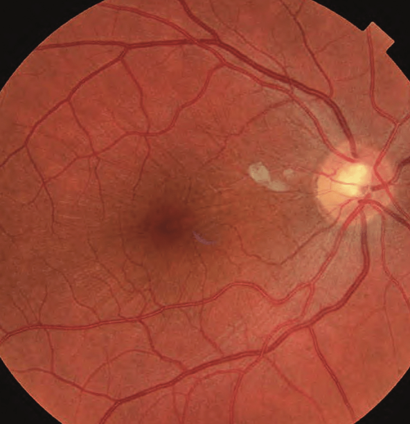
An epiretinal membrane (ERM) is an eye condition where a thin, clear, and transparent fibrous cellular material forms on the surface of the retina. It commonly occurs, affecting the posterior pole of the retina over the macula. The cause is unknown, but some research shows that it can be secondary to trauma, post-intraocular surgery, chronic ocular diseases, and so on.
 Symptoms:
ERMs usually cause several symptoms, such as:
- Difficulty in seeing fine details and recognizing faces when the central part of the retina is affected.
- Blurred and distorted vision.
- Straight lines appear wavy.
- Decreased vision and loss of central vision.
- Double vision.
Causes:
Epiretinal membranes can occur as a result of the normal aging process in the eyes. It is common in individuals over the age of 50. One reason for this condition is when the vitreous gel peels away from the retina. ERM may also form following retinal or eye surgery.
Diagnosis:
An epiretinal membrane can be diagnosed during a routine eye examination for mild cases where the symptoms do not affect the individual. During the eye examination, an optometrist or ophthalmologist may use a non-invasive imaging technique called Optical Coherence Tomography (OCT) to visualize the layers of the retina. In some patients, an ophthalmologist will request additional tests such as fluorescein angiography to determine any other underlying problem that might have caused this problem. The test involves the use of dye to light up areas in the retina.
Treatment:
In many cases, mild membranes are monitored over time for progression. Aside from surgery, there are no other available treatments for epiretinal membranes. Eyeglasses, contact lenses, and prescription eye drops are generally not effective treatments for epiretinal membrane.
Surgery is considered and recommended when the patient’s vision or perception of visual distortion starts to affect their quality of life, for example, when the vision is worse than 20/40 (6/12). To preserve the anatomic integrity of the retina, the membrane removal must be executed with extreme caution. The surgeon will make a tiny cut around 4 mm and remove the fluid from inside the eye. Then, the surgeon will hold and gently peel the epiretinal membrane from the retina and replace the fluid in the eye. The surgery is performed under local anesthesia.
Therefore, it is advisable to visit an ophthalmologist for a detailed eye examination if you experience any unusual symptoms related to an epiretinal membrane (ERM). Moreover, an eye screening can also be beneficial for the early detection of any retinal disorder.
Residents within Klang Valley in areas such as Cheras, Puchong, Shah Alam, Petaling Jaya, and Kepong can visit our low vision specialist, Ms Vinodhini Naidu to have their eyes examined at Nexus Bangsar South KL branch. Residents in Seremban 2, Senawang, Sendayan and Port Dickson can visit Dr Teh Wee Min at our Seremban branch. Residents in Johor Bahru, Skudai, Kulai, Iskandar Puteri, Senai, Tebrau, Batu Pahat, Kluang, Segamat can visit Dr Ling Kiet Phang or Dr Chan Choon Teng at our Johor Bahru branch. Residents in Perai, Bukit Mertajam, Butterworth, Penang island, Alor Setar, Kulim and Sungai Petani can visit Dato’ Dr Haslina at our Penang branch.


